


Varicose veins during pregnancyare venous vasocytes that arise during pregnancy and are pathogenetically related to them. It is manifested by severity, paresthesia, pain in the lower legs and external genitals, swelling, wrinkled muscles, trophic skin lesions. It is diagnosed by examination, ultrasound angioscanning method. During pregnancy, treatment is usually limited to compression therapy with correction of sleep and rest, physical activity, and nutrition. Possible appointment of phlebotonics, phleboprotectors, anticoagulants, antiplatelet agents. Surgical treatment is usually used after childbirth.
General information
Varicose veins (varicose veins) is one of the most common vascular diseases associated with pregnancy. According to the study, up to 15-20% of people suffer from venous pathology, while 2/3 of them are women, and 60-80% of cases of venous ectasia arise as a result of pregnancy. The disease is usually diagnosed for the first time in young patients, 75% of whom are under 30 years old. In more than two-thirds of cases, varicose vein clinics appear after the first 20 weeks of pregnancy. The appropriateness of timely diagnosis of varicose veins is associated with a high probability of fetoplacental insufficiency and the risk of fatal thromboembolic complications in the absence of adequate therapy.
Reason
Taking into account statistical data on the incidence of varicose veins during pregnancy, most experts in the field of obstetrics and gynecology consider this disease as a complication of pregnancy. The predisposing factor that causes vascular ectasia in 91% of patients is genetically determined genetic failure of the central venous sheath, in which the amount of collagen is reduced and the polysaccharide content increases. The development of varicose veins in women with constitutional tendencies during pregnancy is facilitated by:
- Increased circulating blood volume. The increase in BCC in pregnant women ranges from 30-50% (when carrying 1 child) to 45-70% (if there are 2 or more fetuses in the uterus). This compensation mechanism makes it possible to ensure adequate blood supply to the child, the vital organs of the woman and the fetoplacental system.
- Hormonal changes during pregnancy. During pregnancy, the ovaries and placenta intensively release progesterone and relax. Under the influence of this hormone, the smooth muscle fibers of the veins relax, and reconstruction of connective tissue occurs. As a result, the vascular wall overcomes worse with increased intravenous pressure.
- Compression of vessels by pregnant uterus. The growing uterus compresses the inferior vena cava and the iliac vein. Blood flow from the pelvis and lower extremities is disrupted, intravascular pressure increases, which provokes the stretching of the venous wall. The influence of these factors plays an important role in the formation of varicose veins after the 25th week of pregnancy.
- Changes in hemostasis system. As the workforce approaches, blood fibrinolytic activity decreases, and the number of coagulation factors increases. This adaptive mechanism aims to reduce the amount of physiological blood loss during childbirth. This increases the likelihood of pathologically altered vein thrombosis.
An additional etiofactor that contributes to the onset of varicose veins in pregnant women is a decrease in physical activity. With insufficient skeletal muscle work, blood stagnation in the legs and pelvis increases. The condition is exacerbated by the presence of excess weight, where there is an increase in the amount of blood circulating in the patient's vascular bed.
Pathogenesis
The starting point for the development of varicose veins during pregnancy is a disruption of the ability of the venous network valve device. Due to the increased BCC and mechanical blockage to the outflow from the lower part of the leg, when the main vein is squeezed, the blood puts increased pressure on the vascular wall. Genetically inherited connective tissue fiber failure is exacerbated by relaxation of vascular smooth muscle under the action of progesterone. As a result, the venous lumen expands, the valve stops closed, blood is inserted into the vascular system at the bottom of the leg. As the disease progresses, pathological processes can spread to the vulva, vaginal, and small pelvic ring ducts.
Classification
The main criteria for the systematization of the form of varicose veins are the anatomical prevalence of venous stasis and the severity of the disease. This approach allows the selection of different treatment regimens for different variants of the disorder. Taking into account the involvement of various organs in the process, varicose veins in the lower part of the legs, varicose veins of the vulva, varicose veins of the pelvic organs are distinguished. Depending on the severity of the clinical symptoms, the degree of venous expansion in the lower leg is differentiated:
- Compensated varicose veins. There are no external signs of vascular ecstasy, pregnant women record leg fatigue at the end of the day, discomfort in the calf muscles during exercise and brisk walking.
- Subcompensation varicose veins. Vascular patterns ("stars") appear on the skin. In the evening, the legs are swollen, at night there are cramps, numbness, pain. Bruises and scratches heal longer than usual.
- Decomposition varicose veins. Patients are always worried about pain in the legs, swelling is increasing. The veins enlarge significantly, knots. Hyperpigmentation skin. There are signs of eczema and trophic disturbances.
With pelvic varicose veins in pregnant women, the disease also develops gradually. In the first stage, the diameter of the vessel exposed to the pelvic venous plexus does not exceed 5. 0 mm. With the latter, the uterus or ovaries are involved in the process, the lumen of the duct is 6. 0-10. 0 mm. The third is characterized by vein eclasia more than 10 mm with total damage to all pelvic venous plexuses.
Varicose vein symptoms
In 80-82% of patients, the disease appears with a feeling of heaviness, tension, "buzzing" in the legs, increased in the evening and during physical exercise. The symptomatology of varicose veins gradually increases. As the disease progresses in some muscle areas, pain appears, the first developing with long standing, doing physical work. In the worst cases, the pain becomes constant, and the intensity can be pronounced until the pregnant woman has difficulty in free movement. Up to 60% of patients record calf muscle spasms, up to 40-50% - loss of sensitivity, numbness of the legs, up to 30% - itching.
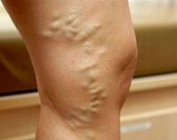
At the subcompensation varicose vein stage, external signs of superficial vein expansion appear. First, areas of the reticular vessels and telangiectasias ("reticules" and "stars") form on the skin. After that, the pattern of veins becomes different. The veins appear dilated, convoluted, eventually nodular. The spread of the ectasia process to the inner canal is evidenced by the occurrence of edema in the ankle and lower leg joints. With the decomposition of varicose veins, the skin of the feet appears hyperpigmented, eczema develops. If the pathology arises long before pregnancy, subcutaneous fat tissue dystrophy, trophic ulcers are possible.
In 4% of patients, the disease affects the veins of the vulva, vagina, and small pelvis. With varicose veins of the vulva and vagina, discomfort, distension, heaviness, itching are observed in the external genital area. There may be swelling of the perineum and labia, bleeding contact from the vagina after sexual intercourse. Pelvic congestion syndrome is manifested by pulling or pain in the lower abdomen, which radiates to the lower back, sacrum, groin, and external genitalia. Dyspareunia (pain during intercourse) is characteristic. In severe cases, dissected disturbances are detected.
Complications
In the absence of adequate treatment, varicose veins in pregnant women can be complicated by the development of trophic ulcers, eripelas, thrombophlebitis, superficial and deep vein thrombosis, pulmonary artery thromboembolism and other large vessels during childbirth. In 40-45% of cases, placental insufficiency occurs with acute and chronic fetal hypoxia. In 25% of patients, observed labor abnormalities (labor weakness, irregular contractile activity of the myometrium) With vaginal varicose veins, major postpartum trauma is possible. Nearly one-third of women who give birth experience defects in placental separation and placental abruption. The long-term consequences of varicose veins that occur during pregnancy are hemorrhoids, paralysis of chronic venous insufficiency, and pelvic pain.
Diagnostics
With the appearance of special skin symptoms, the diagnosis of varicose veins during pregnancy usually does not cause any difficulties. The task of the diagnostic level is to determine the degree and localization of venous ectasia, to exclude other causes that can cause stagnation in the blood vessels in the lower leg. The most informative survey methods are:
- Chair Check. This study revealed characteristic changes in the veins in the vulvar area and inner thighs - ectasia, tortuosity, nodosity. Swelling of the labia and perineum is possible. When viewed in a mirror, the vaginal mucosa appears hypertrophic, cyanotic. Vaginal arch with bimanual palpation smoothed, often painful.
- USDG venous system. During the ultrasound scan, the shape and diameter of the vessel, its length, anatomical position, and wall condition were assessed. This method makes it possible to determine the branching zone, valve tool consistency, vein patency, presence and direction of reflux. You can scan both the vessels at the bottom and the inferior vena cava (ultrasound IVC).
- Duplex duplex scanning. The advantage of the non-invasive method, which combines traditional ultrasound and Doppler studies, is not only obtaining detailed information on blood flow parameters, but also visualization of venous tissue. Duplex angioscanning is used for a comprehensive assessment of the condition of the superficial, perforated and deep vessels.
Radiodiagnostic methods (varicography, selective ovarycography, ascend phlebography of the extreme, pelvic phlebography, CT venography, phleboscintigraphy, etc. ) during pregnancy are used to a limited extent due to possible negative effects on the fetus. In difficult cases, with suspicion of pelvic varicose veins, diagnostic laparoscopy is performed with caution. Differentiation of leg varicose veins is made with falling pregnant women, heart failure, lymphedema, acute thrombosis of the venous system. Small pelvic varicose veins must be distinguished from genital endometriosis, chronic inflammatory pathology of the pelvic organs, submucosal and lower uterine myoma, cysts and other ovarian tumors. In addition to the observations of obstetricians-gynecologists, patients are recommended to consult a phlebologist, cardiologist, and oncologist.
Treatment of varicose veins during pregnancy
The main objective of therapy for varicose veins in pregnant women is to stop the development of the disorder, reduce the severity of the clinical picture and prevent possible complications of thromboembolism. Non-pharmacological methods are considered better, if necessary in addition to pharmacotherapy during a safe pregnancy:
- Compression therapy. A woman with a confirmed diagnosis of varicose veins is recommended to wear it every day throughout pregnancy, to use elastic bandages, compression tights or stockings of 1-2 compression classes during childbirth and the postpartum period. Compression treatment by mechanically reducing the diameter of the superficial veins speeds up blood flow, reduces swelling and congestion.
- Herbal flebotonics and phleboprotectors. The effects of the use of drugs in this group are associated with increased venous wall tone, decreased permeability, increased microcirculation, rheological properties of blood and lymph outflow. The advantage of most bioflavonoids is that they can be used during pregnancy and lactation. Phlebotonic drugs are prescribed in tablet and external form.
- Anticoagulants and antiplatelet agents. With signs indicating an increased tendency to clot and the threat of DIC expansion, drugs with antithrombotic activity are used with caution. To improve blood rheology and vascular microcirculation, pharmaceutical agents are indicated that inhibit platelet accumulation and have an angioprotective effect.
Pregnant women with varicose veins are recommended a complex of physiotherapy exercises, lymphatic drainage massage, walking, daily contrast bath. Diet correction involves the intake of foods rich in fiber and vegetable fats. Injection sclerotherapy, miniflebectomy, crossectomy, endovasal laser coagulation and other surgical treatment methods are used in rare cases with severe forms of the disease, severe pain syndrome, and the presence of complications. Often, surgical correction is performed at the end of the breastfeeding period.
Delivery tactics
The preferred method of birth for varicose veins is natural birth, initially elastic bandages or compression garments are used on the lower limbs of women giving birth. Patients with vulvar-vaginal varicose veins require careful treatment over a continuous period with the implementation of protective perineotomy according to the instructions. When a ruptured vein ruptures, the damaged duct is carefully tied with repeated sutures from the node conglomerate. Caesarean section is recommended for patients at high risk of complications of thromboembolic and severe varicose veins.
Prediction and prevention
With timely detection and adequate therapy, the prognosis is better. For prophylactic purposes, it is recommended to get a good night's sleep and rest periodically throughout the day in a supine position with the feet placed on a firm surface at an angle of 30 °. Pregnant women with burdensome offspring should refuse to wear shoes with heels over 5 cm, limit sitting or standing periods, and control weight gain.
To prevent varicose veins, walking every day, reducing salt intake, taking vitamin preparations that strengthen the vascular wall are effective. Patients with varicose veins planning a pregnancy, according to the indications, undergo surgical intervention to correct the disease.